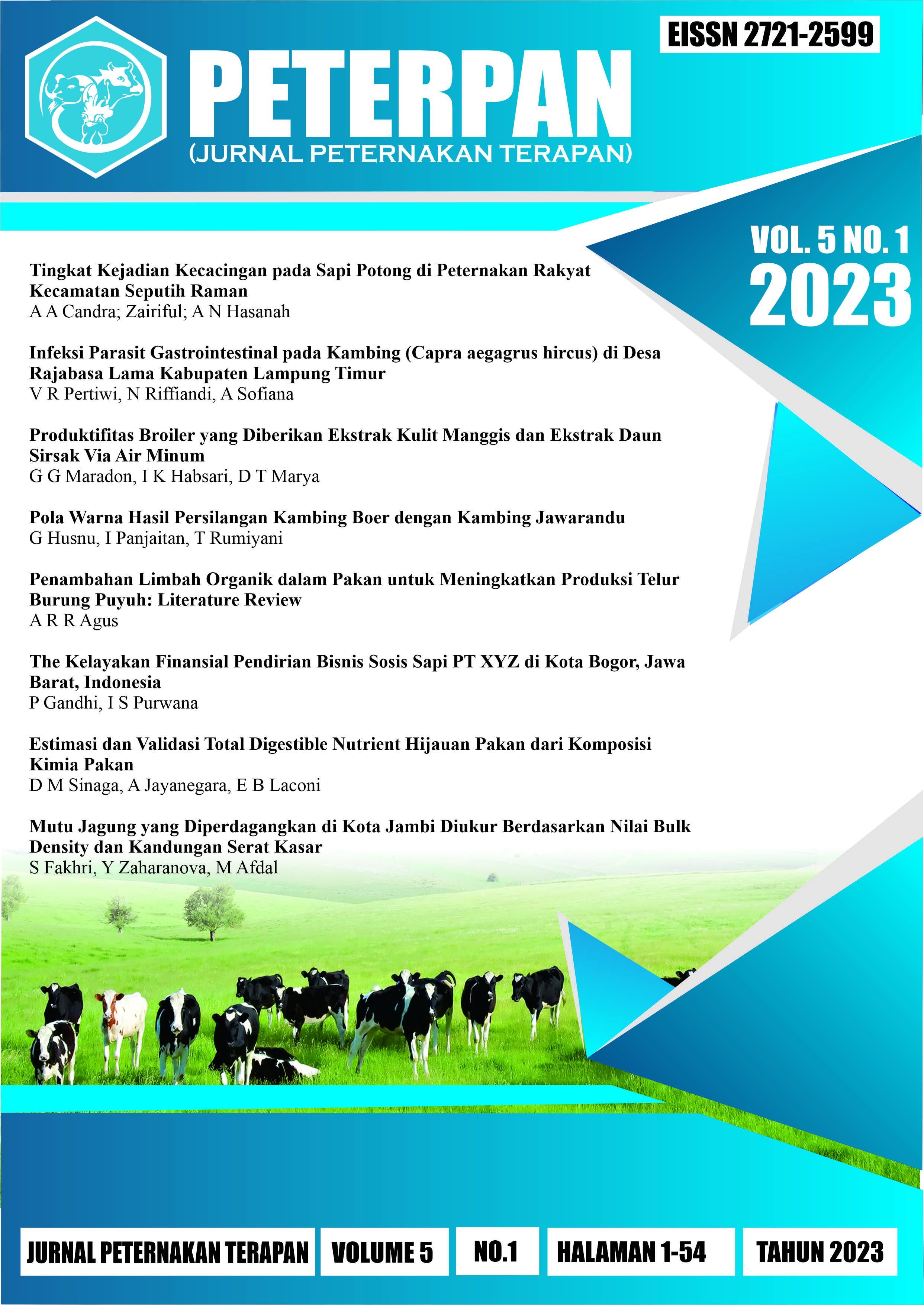
Infeksi Parasit Gastrointestinal pada Kambing (Capra aegagrus hircus) di Desa Rajabasa Lama Kabupaten Lampung Timur
DOI:
https://doi.org/10.25181/peterpan.v5i1.2829Keywords:
gastrointestinal, goat, infection, parasiteAbstract
In developing countries such as Indonesia, the health of small ruminants such as goats is not given much attention because the medical costs are very high, it causing a farmer to prefer to sell their livestock, even though at relatively low prices if there are signs of infection, one of which is due to parasitic diseases. This research was carried out in the village of Rajabasa Lama. The study was conducted using a descriptive method by collecting feces from the goat pens in that area using native methode and sugar floatation method Furthermore, the examination was carried out using a native test and fecal floating examination using a fluid sugar medium. The results of the examination of gastrointestinal tract parasites that were found included parasites from the protozoan Entamoeba sp. and Eimeria sp. and also parasites from the Trematoda family, the eggs of the worm Fasciola sp.. Eimeria sp. is a parasite that quite often infects ruminants, including goats. This study showed that goats in Rajabasa Lama Village had gastrointestinal parasite infections including Eimeria sp., Entamoeba sp., Fasciola sp. worm eggs, and Trichuris sp. eggs.Downloads
References
Andrews, A. 2022. Coccidiosis of Goats - Digestive System - MSD Veterinary Manual
Anuracpreeda P, Wanichanon C, dan Sobhon P. 2008. Paramphistomum cervi antigenic profile adults as recognized by infected cattle sera. Exp Parasitol 118:203–207
Arsenopoulos, K.V., Fthenakis, G.C., Katsarou, E.I. and Papadopoulos, E. 2021. Haemonchosis: A challenging parasitic infection of sheep and goats. Animals, 11(2), p.363. https://doi.org/10.3390/ani11020363
Awaludin, A. and Nusantoro, S. 2018. Identify the diversity of helminth parasites in cattle in Jember district (East Java-Indonesia). IOP Conference Series: Earth and Environmental Science 207(1): 012032. IOP Publishing.
Bulbul, K.H., Akand, A.H., Hussain, J., Parbin, S. and Hasin, D. 2020. A brief understanding of Trichuris ovis in ruminants. International Journal of Veterinary Sciences and Animal Husbandry 5(3): 72-74
Chali AR, Hunde FT. 2021. Study on prevalence of major gastrointestinal nematodes of sheep in Wayu Tuka and Diga District, Oromia Regional State. Vet Med Open J. 2021; 6(1): 13-21. doi: 10.17140/VMOJ-6-154
Hing S, Othman N, Nathan SKSS, Fox M, Fisher M, and Goossens B. 2013. First Parasitological Survey of Endangered Bornean Elephants Elephas maximus borneensis. Endangered Species Researh. 21 : 223- 230.
Horak IG and Clark R. 2000. Studies on Paramphistomiasis. V. The pathological physiology of the acute disease in sheep. Onderstepoort J Vet Res 30:145–160
Jacobson, C., Williams, A., Yang, R., Ryan, U., Carmichael, I., Campbell, A.J., and Gardner, G.E. 2016. Greater intensity and frequency of Cryptosporidium and Giardia oocyst shedding beyond the neonatal period is associated with reductions in growth, carcase weight and dressing efficiency in sheep. Vet. Parasitol. 228, 42–51.
Rolfe F, Boray JC, and Collinis G.H. 1994. Pathology of infection with Paramphistomum ichikawai in sheep. Int J Parasitol 24:995–1004
Singh RP, Sahai BN, and Jha GJ .1984. Histopathology of the duodenum and rumen during experimental infections with Paramphistomum cervi. Vet Parasitol 15:39–46
Skirnisson, K., and Hansson, H.. 2006. Causes of diarrhoea in lambs during autumn and early winter in an Icelandic flock of sheep. Icel. Agric. Sci. 19, 43–57.
Stensvold, C.R., Lebbad, M., Clark, C.G., 2010. Genetic characterisation of uninucleated cyst-producing Entamoeba spp. from ruminants. Int. J. Parasitol. 40, 775–778.
Suandhika, P., Dwinata, I.M. and Arjana, A.A.G. 2017. Prevalensi Nematoda gastrointestinal pada gajah sumatera di Bakas Elephant Tour dan Taro Elephant Safari Park. Indonesia Medicus Veterinus 6(3): 213-221. DOI: 10.19087/imv.2017.6.3. 213
Taylor, M.A.; Coop, R.L.; and Wall, R.L. 2015.Veterinary Parasitology, 4rd ed.; Blackwell Publishing: London, UK
Tehrani, A., Javanbakht, J., Khani, F., Hassan, M.A., Khadivar, F., Dadashi, F., Alimohammadi, S. and Amani, A., 2015. Prevalence and pathological study of Paramphistomum infection in the small intestine of slaughtered ovine. Journal of parasitic diseases, 39, pp.100-106.
Tolistiawaty, I., Widjaja, J., Lobo, L.T. and Isnawati, R., 2016. Parasit gastrointestinal pada hewan ternak di tempat pemotongan hewan Kabupaten Sigi, Sulawesi Tengah. Balaba: Jurnal Litbang Pengendalian Penyakit Bersumber Binatang Banjarnegara 12(2) :71-78.
Woodbury, M.R., Copeland, S., Wagner, B., Fernando, C., Hill, J.E. and Clemence, C., 2012. Toxocara vitulorum in a bison (Bison bison) herd from western Canada. The Canadian Veterinary Journal. 53(7):791-794.
Downloads
Published
How to Cite
Issue
Section
License
Copyright (c) 2023 PETERPAN (Jurnal Peternakan Terapan)

This work is licensed under a Creative Commons Attribution-NonCommercial-ShareAlike 4.0 International License.


 Under Licenced by